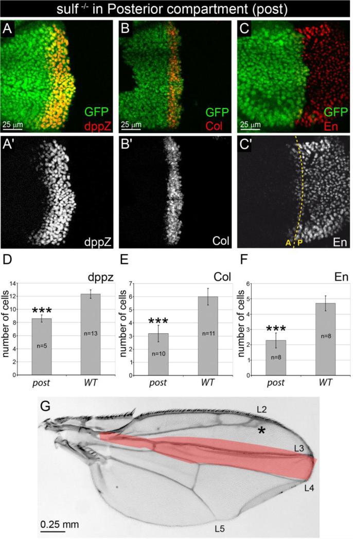
Figure 3. Removal of DSulf1 activity in Hh producing posterior compartment results in Hh loss-of-function phenotype.
(A-C’) Expression patterns of dppZ (A, A’), Col (B, B’) and En (C, C’) in Dsulf1ΔP1 mitotic clone restricted to the P compartment, detected by absence of GFP staining (green). (D-F) Number of cell rows expressing dppZ (D), Col (E) and En (F) in wing discs containing posterior (post) Dsulf1ΔP1 clones compared to wt. Note the narrowing of dppZ, Col and anterior En expression domains compared to wt. Error bars represent the standard deviation (***=P<0.0005 using a t-test). (G) Adult wing carrying posterior mutant clones showing a narrowing of the L3-L4 spacing (21±2 trichomes), compared to wt (30±1 trichomes, red in G). Note that positioning of the L2 vein is also affected (asterisk).

