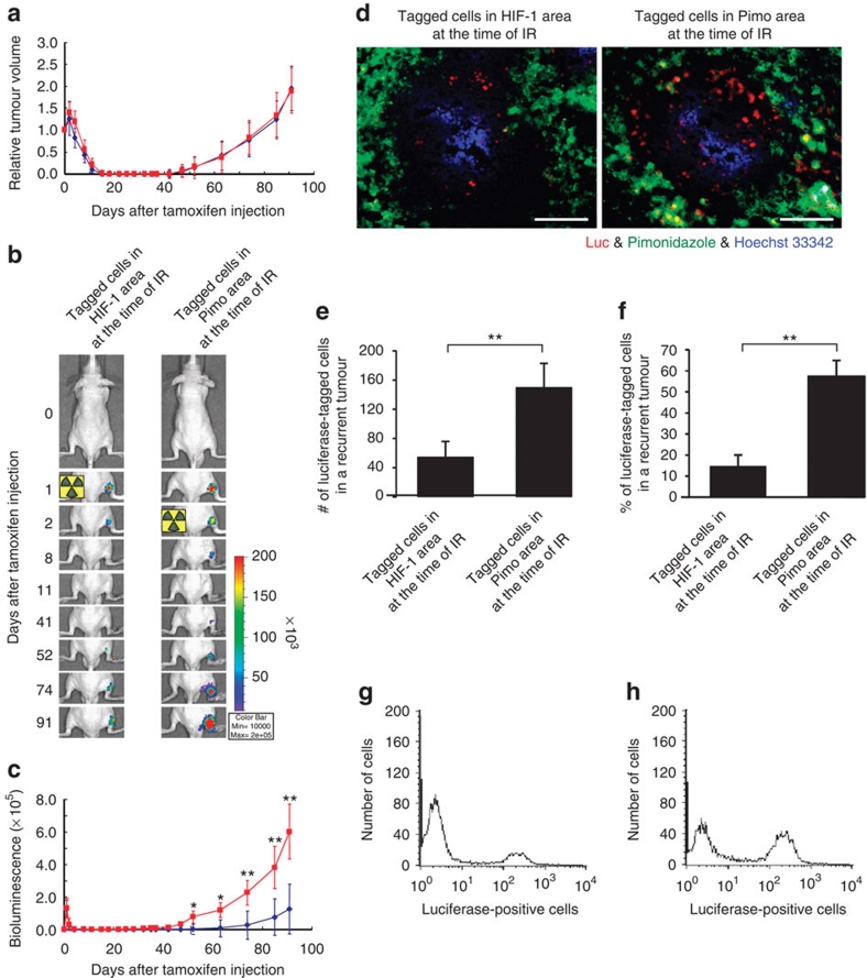
Figure 4. Fate of hypoxic tumour cells after radiation treatment.
(a) Tumour-bearing mice were γ-irradiated on either the first day after tamoxifen treatment when tagged cells were located in the HIF-1-positive cell area (blue rhombus; group name: tagged Cells in HIF-1 area), or the second day after tamoxifen treatment when the tagged cells were located in the pimonidazole-positive cell area (red square; group name: tagged cells in Pimo area). Relative tumour volume was calculated as the ratio of the tumour volume on each day to the corresponding volume on day 0. Mean±s.d. n=12. (b) During the experiment in (a), tumour-bearing mice were subjected to optical imaging at the indicated time points. Representative images are shown. (c) Bioluminescence intensities were quantified based on the optical imaging in (b). Blue rhombus: group of 'tagged cells in HIF-1 Area'. Red square: group of 'tagged Cells in Pimo area'. Mean±s.d. n=12. *P<0.05. **P<0.01 (Student's t-test). (d) The tumour xenografts in each group of the experiments (a–c) were surgically excised 91-days post-injection of tamoxifen and cut into two pieces. One of them was subjected to immunohistochemistry with anti-luciferase (red) and anti-pimonidazole (green) antibodies. Hoechst 33342: perfusion marker (blue). Bar, 50 μm. Reproducibility was confirmed in 40 tumour cords in 12 independent sections. (e) The number of luciferase-positive cells per tumour cord was counted in the tumour sections in (d). Mean±s.d. n=40 tumour cords in 12 independent sections. **P<0.01 (Student's t-test). (f) Single cell suspension was prepared from another piece of xenografts in (d) and subjected to flow cytometric analysis with anti-luciferase antibody to quantify percentage of luciferase-positive cells in the recurrent tumour of each group. Mean±s.d. n=12 independent tumour xenografts. **P<0.01 (Student's t-test). (g) Representative flow cytometric histogram in the 'tagged cells in HIF-1 area group'. Reproducibility was confirmed by using 12 independent xenografts. (h) Representative flow cytometric histogram in the 'tagged cells in Pimo area group'. Reproducibility was confirmed by using 12 independent xenografts.

