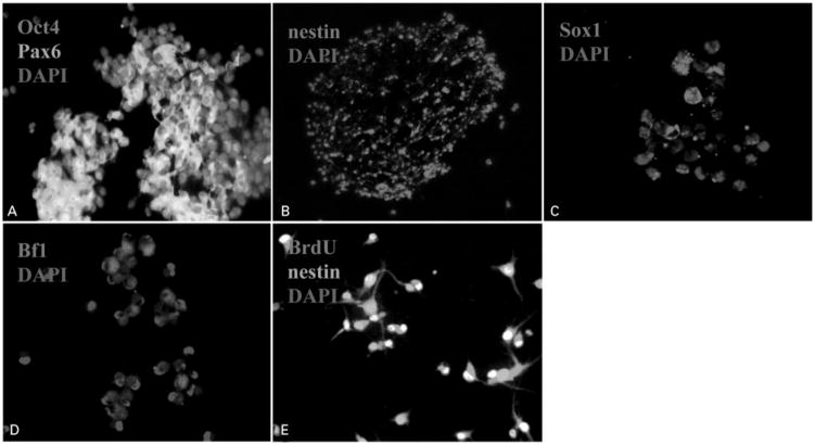
Figure 1.
Micrographs showing the production of neural stem cell (NSC)-like cells from bone marrow (BM)-derived human mesenchymal stem cells (hMSCs). (A) Octamer-binding protein 4 (Oct4) (red) and paired box gene 6 (Pax6) (green)-immunoreactive cells in the cellular aggregates of hMSCs (×20). Note that some Oct4-immunoreactive cells colocalized with Pax6-immunoreactive cells. (B) Nestin-immunoreactive cells (red) in the neurosphere-like structures (×10). (C) Bf1 (red)-immunoreactive cells in the NSC-like cells (×20). (D) Sox1 (red)-immunoreactive cells in the NSC-like cells (×20). (E) Bromodeoxyuridine (BrdU) (red) and nestin (green)-immunopositive cells (×20). Note that most of the nuclei of nestin-immunopositive cells were labeled by BrdU, suggesting that these cells are dividing cells. Cell nuclei were counterstained with 4′,6′-diamidino-2-phenylindole (DAPI) (blue).

