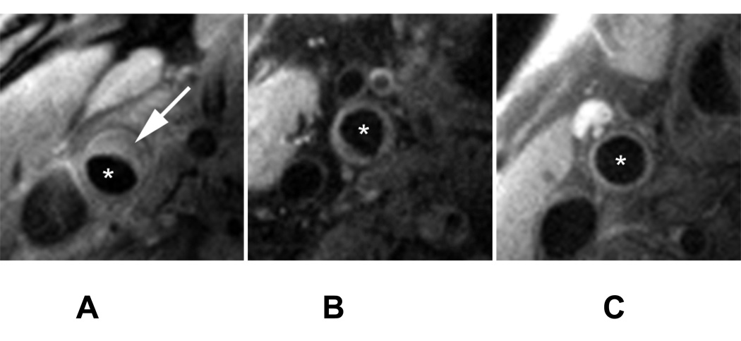
Figure 2.
MR Images of the Common Carotid of subjects with atherosclerosis. A: mean wall area: 39 mm2, B: mean wall area: 30 mm2, C: mean wall area: 17 mm2. Arrow points to atherosclerotic plaque and asterisk indicate the lumen. The imaging parameters were as follows: proton density weighted (PDW) non-gated sequence imaging 12 slices simultaneously (TR/TE = 2130/5.6 ms), with a field of view of 12 × 12 cm, bandwidth of 488 Hz/pixel, matrix size of 256 × 256, a turbo factor of 15 and 2 signal averages. A chemical shift suppression pulse was used to suppress signal from perivascular fat, not affecting the signal from intraplaque lipids.

