
Figure 2.
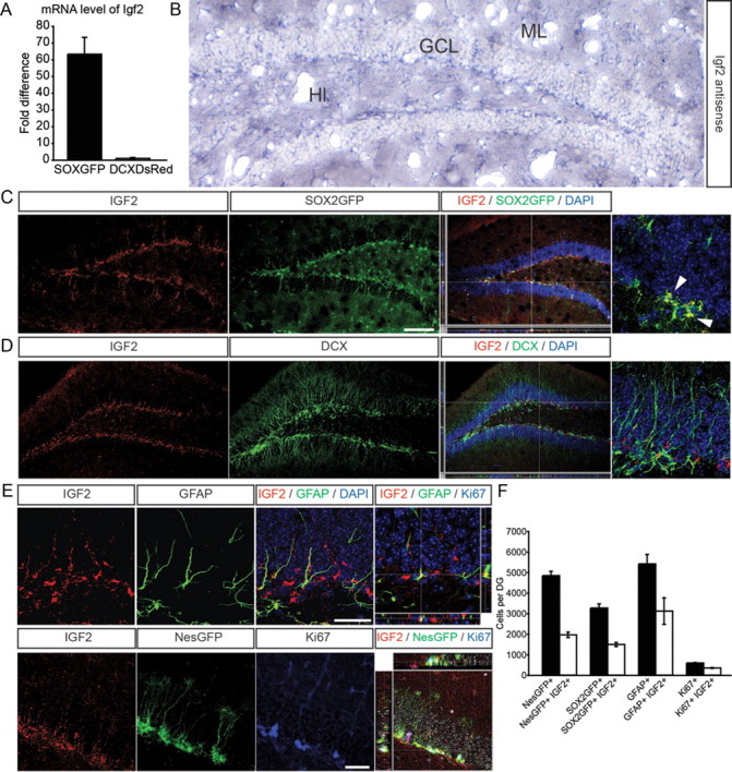
IGF2 is expressed in the SGZ of the DG. A, Quantitative RT-PCR shows strong enrichment of Igf2 in SOX2+ cells of the DG compared with DCX+ cells. B, In situ hybridization using a riboprobe against Igf2 mRNA shows expression in the granule cell layer (GCL) and an enrichment of Igf2 mRNA in the SGZ of the DG. Hl, hilus; ML, molecular layer. C, IGF2 (red) shows high expression in SOX2+ cells (green) in the DG. A high-power view of a SOX2GFP/IGF2 coexpressing cell is shown on the far right (arrowheads). D, IGF2 (red) shows only low expression in DCX+ cells (green). A high-power view of a DCX+ cell (green) that does not colabel with IGF2 is shown on the far right. E, Representative images of IGF2 expression (red) in costainings with GFAP (top, green), NestinGFP (bottom, green), and Ki67 (bottom, blue). Z-stacks show 3-dimensional reconstruction of IGF2/GFAP colabeled cells (top) and IGF2/NestinGFP colabeled cells (bottom). F, Using confocal microscopy, we quantified the number of NestinGFP+, SOX2GFP+, GFAP+, and Ki67+ cells colabeling with IGF2. We found that a substantial fraction of radial-glia like cells in the DG expressed IGF2. Nuclei were counterstained with DAPI. Data are presented as mean ± SEM. Scale bars (in C) C, D, 100 μm; E, 50 μm.