Figure 6.
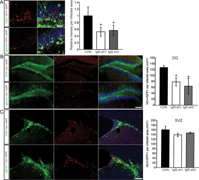
IGF2 knockdown reduces NSC proliferation in the adult DG. A, Fluorescence intensity measurements of IGF2 staining in either nontargeting shRNA or shRNA against Igf2-infected cells. Shown are confocal images of the DG after injection of a control virus expressing a nontargeting shRNA (CON, green, top) or virus expressing shRNA against Igf2 (Igf2-sh1, green, bottom) and stained for IGF2 (red, left). Quantification of the IGF2 staining in virus-transfected cells of the DG, showing a reduction in the relative fluorescence level in the Igf2 knockdown cells compared with the CON shRNA cells. B, Shown are confocal images of the DG after injection of a control virus expressing a nontargeting shRNA (green, top) or virus expressing shRNA against Igf2 (Igf2-sh1, green, bottom). Proliferating cells in the DG were visualized by IdU labeling (red). Note the reduction in IdU-labeled cells in the DG. Right, an overlay of single channels. C, Shown are confocal images of the SVZ after injection of a control virus (green, top) or virus-expressing shRNA against Igf2 (Igf2-sh1, green, bottom). Right, an overlay of single channels. In contrast to the DG, IGF2 knockdown does not impair proliferation in the SVZ. Data are presented as mean ± SEM. Scale bars, 100 μm. *p < 0.05.

