
Figure 2.
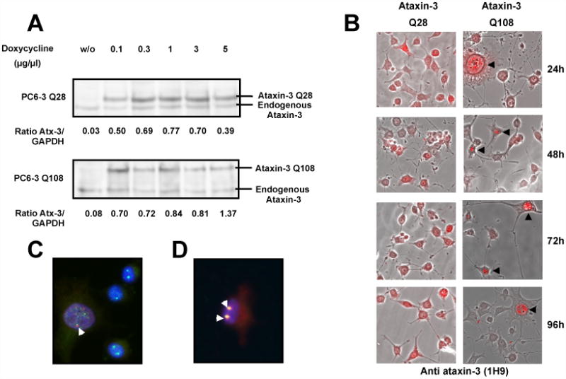
Aggregates of human expanded ataxin-3 in PC6-3 ataxin-3 Q108 cells are primarily nuclear. (A) Total extracts were prepared from PC6-3 ataxin-3 Q28 cells or PC6-3 ataxin-3 Q108 cells incubated in the absence or presence of increasing concentrations (0.1, 0.3, 1, 3 and 5 μg/μl) of doxycycline for 24 hours and subsequently probed for ataxin-3 on a western blot. (B) Merged representative images of fluorescent and optical differential interference contrast microscopy of PC6-3 cells incubated with doxycycline (1 μg/μl) for expression of human ataxin-3 Q28 or human ataxin-3 Q108 during 24, 48, 72 or 96 hours and immunostained for ataxin-3 (in red). (C, D) Representative images of PC6-3 ataxin-3 Q108 cells expressing human expanded ataxin-3 for 48 hours, which were fixed and immunostained for ataxin-3 (in red) and coilin (in green) (C) or PML protein (in green) (D). Yellow shows co-localization between proteins. Nuclei were stained with Hoechst 33342 (1 μg/ml).