
Figure 3.
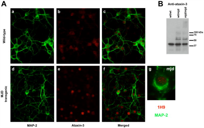
Aggregation in the human expanded ataxin-3 transgenic mouse. (A) Cerebellar granule cells were isolated from 7 days wild-type (a, b, c) or transgenic (d, e, f) pups and kept in culture for 7 days. Cells were stained for MAP-2 (a, c, d, f - in green) and ataxin-3 (b, c, e, f - in red) and visualised through fluorescence microscopy. (g) Higher magnification of transgenic cerebellar granule cell immunostained for MAP-2 (in green) and ataxin-3 (in red). (B) Western blotting for ataxin-3 was performed in total brain extracts of wild-type (wt/wt), or transgenic (one copy (wt/mjd) or two copies (mjd/mjd) of the transgene) 4 month old mice to evaluate the expression of the transgene and the aggregation of human expanded ataxin-3.