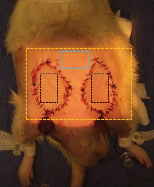
Fig. 1.

Diagram of typical orientation of the flaps on the animal and the region imaged by the prototype spatial frequency domain imaging device (Tissue OxImager). The illumined region out-lined by the golden box is the region imaged by the device. The blue box is the area outside of the surgical field used as the native skin group. The black boxes represent the area on either the control flap or experimental flaps (selective arterial or selective venous occlusion flaps). Note the incisions caudal to the flaps used to perform selective ligation of the vessels.
