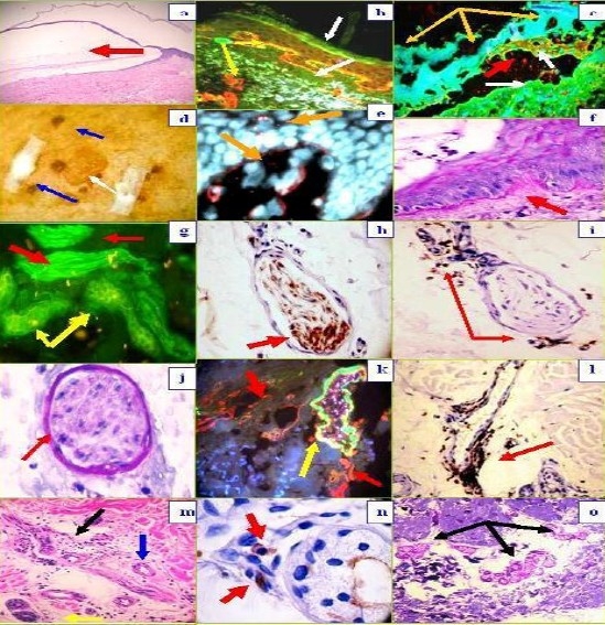
Fig. 1a.

H & E staining (100X) demonstrating the subepidermal blister (red arrow). 1b, DIF, positive staining with FITC conjugated anti-human IgE at the BMZ in a linear pattern (green staining, lower white arrow), colocalizing with the collagen IV(CIV) antibody (red staining, top yellow arrow). Please notice that CIV also stains the upper dermal blood vessel areas (lower yellow arrow). The epidermal corneal layer also shows some IgE reactivity (upper white arrow). 1c. DIF, showing destruction of epidermal keratinocytes, characterized by amorphous staining of cell nuclei with Dapi (blue staining, yellow arrows). The red arrow shows defragmented pieces of CIV in the blister lumen (red particles); the white arrows show positive staining with FITC conjugated antihuman fibrinogen, on both sides of the blister (epidermal and dermal). 1d , Shows a clinical blister (white arrow) and some adjacent crusts (blue arrows). 1e, DIF. Note the keratinocytes nuclei in blue (Dapi), and the mapping of the blister with CIV antibody (red staining, yellow arrows). Please note the delicate, trans-epidermal excretion of tiny fragments of CIV, seen as small red dots among the keratinocytes. 1f. Periodic acid-Schiff (PAS) stain, displaying pink positivity at the BMZ (red arrow). 1g. Positive staining of a nerve with FITC conjugated anti-human fibrinogen (green staining; red arrows), as well as against eccrine sweat glands (yellow arrows). 1h. The identity of the nerve was confirmed by colocalizing, positive S-100 IHC staining (brown staining, red arrow). 1i, 1l and 1n. In addition, we utilized IHC to confirm a significant lymphocytic infiltrate around dermal neurovascular package and eccrine sweat glands with CD45 (brown staining, red arrows) . 1j. PAS positive, pink staining of a nerve sheath (red arrow). 1k, DIF positive staining of a blood vessel wall with FITC conjugated Complement/C3 (green-yellowish staining, yellow arrow), colocalizing with CIV antibody (red staining, red arrows). 1m. H & E stain, showing lymphocytes infiltrating around dermal nerves (black arrow), eccrine sweat glands (yellow arrow) and an eccrine sweat gland ductus (blue arrow). 1o. PAS positive staining of eccrine sweat gland peripheral membranes (pink staining, black arrows).
