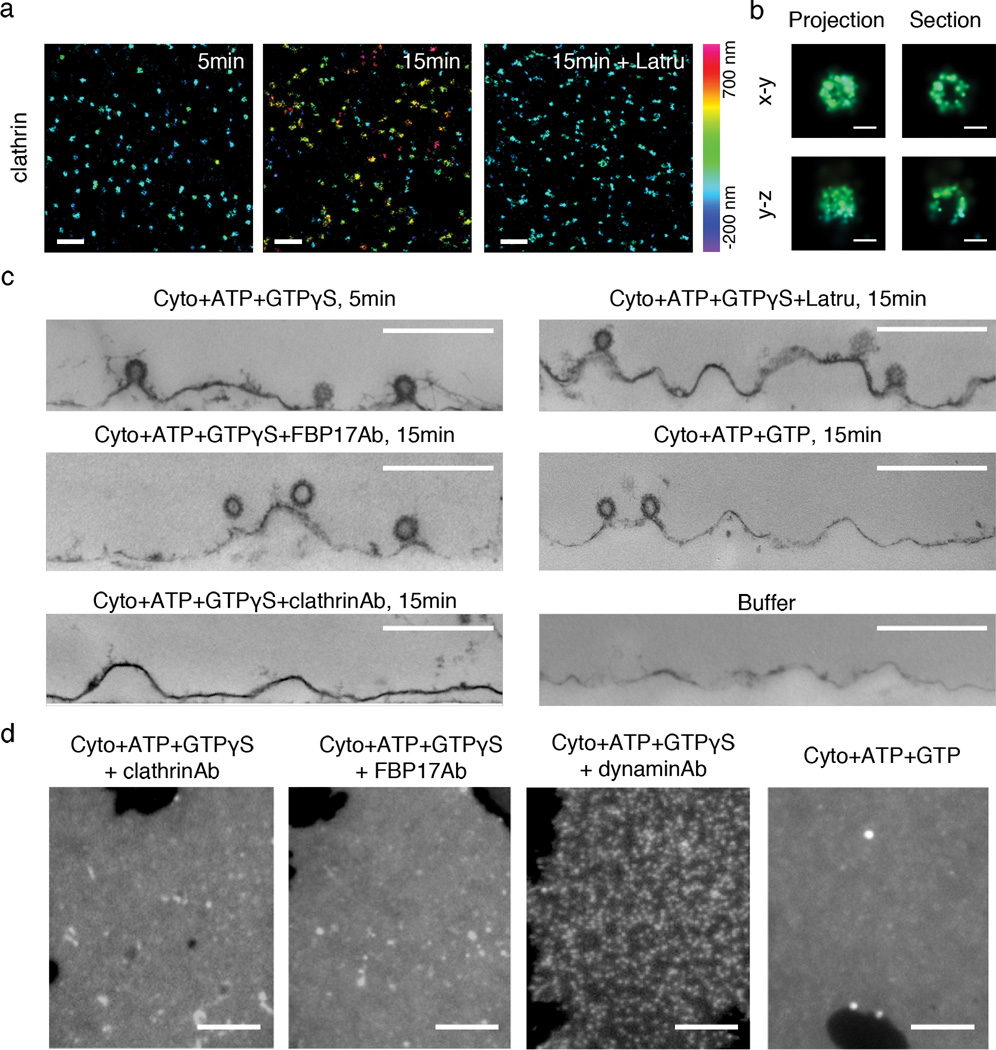
Figure 3.
Tubular invaginations require spatially coordinated action of clathrin, actin and FBP17. (a) 3D-STORM images of clathrin (Alexa 405-Cy5 labeled clathrin heavy chain) at different time points after incubation with cytosol, ATP plus GTPγS and in the absence or presence of latrunculin B (Latru). Two color 3D imaging was performed together with PM-GFP (not displayed here). (b) High-magnification view of 3D-STORM images of a single clathrin-coated pit. In the right panels, the horizontal and vertical projections of the 3D image of one clathrin structure display the half-spherical shape of the pit, whereas 50 nm thick cross sections through the geometric center of the pit reveals its hollow inside (in both x–y and y–z views) and the opening at the bottom (only in y–z view), as expected for clathrin-coated pits. In both (a) and (b), color of individual points in the images encodes distance from the average height of the basal membrane sheet, ranging from -200 nm to 700 nm. (c) Thin section EM confirms the presence of clathrin-coated pits but a lack of tubulation at early point (5min) of incubation with cytosol, ATP plus GTPγS, or at later time point (15min) in the presence of latrunculin B, anti-FBP17 antibodies (FBP17Ab), or GTP instead of GTPγS. In the absence of cytosol or in the presence of anti-clathrin antibodies (clathrinAb), clathrin-coated pits are rare. (d) Fluorescence images of plasma membrane sheets (labeled by PM-GFP) after incubations as indicated. Scale bars: 1 µm (a); 100 nm (b); 500 nm (c); 5 µm (d).

