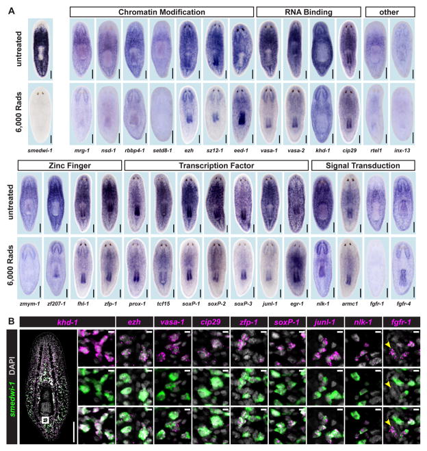
Figure 2. Irradiation-Sensitive Transcripts are Expressed in smedwi-1+ Proliferating Cells.
(A) Whole-mount in situ hybridization (ISH) in untreated animals and animals fixed 5 days after 6,000 Rads γ-irradiation. Genes were annotated by BLASTx and PFAM (See also Supplemental Table 3). (B) Expression of genes identified by microarray, analyzed by double FISH with a smedwi-1 RNA probe (proliferative cell marker). Zoomed images are single confocal planes from tail regions. Most cells detected by FISH co-expressed smedwi-1; cells with little/no smedwi-1 gene expression are labeled by arrowheads. Some transcripts (e.g., ezh and cip29) are expressed at low levels with background signal (scattered magenta dots) also visible. See also Supplemental Figure 2. Shown are representative ventral views, anterior up. Scale bars 200 μm, 10 μm (zoomed images).

