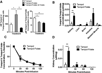
Figure 2.
Tempol-folate is selectively taken up by cultured HK-2 cells as well as folate receptor-rich mouse tissues and remains detectable in the plasma up to 120 minutes postinfusion. (A) Concentration of tempol and tempol-folate in HK-2 cells resuspended in KHB and media, quantified by ESR after 1-hour incubation with 1 μM tempol, tempol-folate, or tempol-folate with 1 μM folate. *P<0.05 and **P<0.01 comparing tempol with other groups; †P<0.05 comparing tempol-folate with combined tempol-folate and folate treatment (n=3 per group). (B) Relative tissue tempol and tempol-folate concentration quantified by ESR in mice after 48-hour intravenous infusion at 500 μg/kg per h. *P<0.05 between tempol and tempol-folate concentration (n=4–7). (C) Postinfusion tempol and tempol-folate plasma concentration measured by ESR. (D) Tempol and tempol-folate concentration in kidneys from mice receiving intravenous infusion of tempol or tempol-folate for 48 hours before withdrawal of infusion. **P<0.01 comparing tempol and tempol-folate concentration (n=2–11 per group). Data represent mean ± SEM.

