Figure 3.
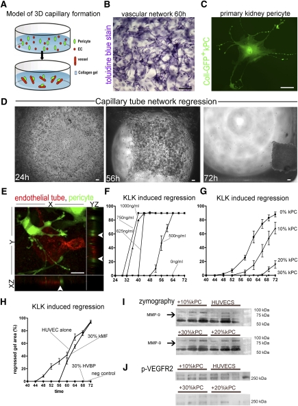
Kidney pericytes stabilize capillary tubes in a 3D gel assay. (A) Schema showing the addition of ECs (red) to gel in wells that spontaneously form capillary networks. Addition of pericytes to this assay permits migration and binding of pericytes to capillary tubes. (B) Toluidine blue–stained gel showing capillary tube network (ECs only) within the gel (bar=100 μm). (C) Kidney pericyte (GFP+) in culture (bar=25 μm). Note cell processes extending the length of several cell bodies. (D) Low-power light images of gels containing capillary tubes (ECs only). Under the influence of the coagulation cascade serine protease, KLK, endothelial tubes are destabilized in the gel and vessels become disorganized leading to progressive collapse of the gel (bar=100 μm). (E) Confocal image of 3D gel with YZ and XZ stacks showing capillary tube (red, CD34) and kidney pericyte (green, Coll-GFP). Note the attachment of kidney pericytes to the capillary tube and numerous processes attached to the capillary tube. Z stacks (arrowheads) show yellow color at points of direct interaction of pericyte processes with capillary tubes, suggestive of peg and socket junctions (bar=25 μm). (F) Dose-response curves measuring collapse of collagen gel (ECs only) induced by KLK. (G) Gel collapse curves induced by KLK (625 ng/ml) in the presence of increasing numbers of kidney pericytes. Note 30% kidney pericytes completely prevent gel collapse (n=12–16/timepoint). (H) Gel collapse curves in response to KLK (625 ng/ml) in the presence of 30% human vascular brain pericytes (HVBPs) or 30% mouse kidney myofibroblasts (kMF). (I) Gelatin zymography of supernatants from collagen gels in the presence of KLK showing multiple bands of gelatinase activity. Note that an approximately 72-kD band of activity, representing MMP9, is completely lost by the addition of 20% or 30% pericytes and the major approximately 60-kD band of MMP2 is attenuated by the presence of 30% pericytes. (J) Phosphoblot for proteins from the collagen gels of KLK-activated gels detecting phospho-VEGFR2. Note that kidney pericytes complete suppress signaling at VEGFR2.

