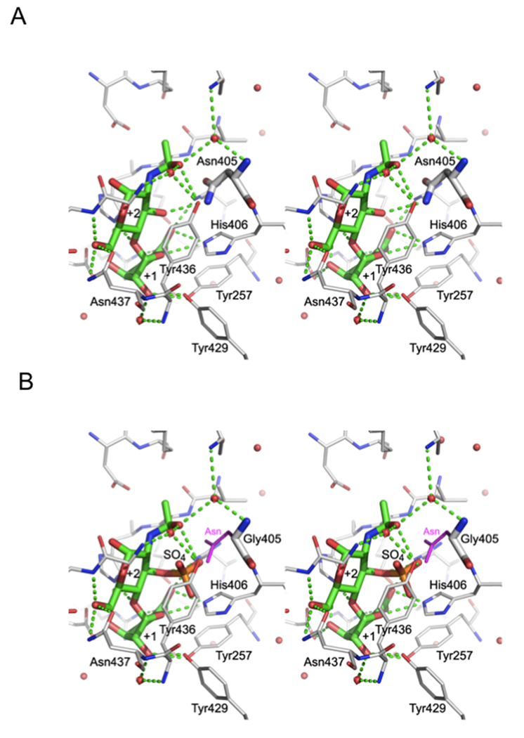
Figure 2.
Vicinity of the +2 sugar. A. The stereo view of crystal structure of WT HepII with bound disaccharide [13] occupying subsites +1 and +2. The disaccharide and Asn405 are shown in thick lines while the neighboring sidechains are in thinner lines. B. The N405G mutant was modeled with a disaccharide containing a 3-O-sulfo glucosamine residue at subsite +2. The asparagine sidechain of the wild type enzyme is painted in magenta to show that it would collide with the 3-O-sulfo group.

