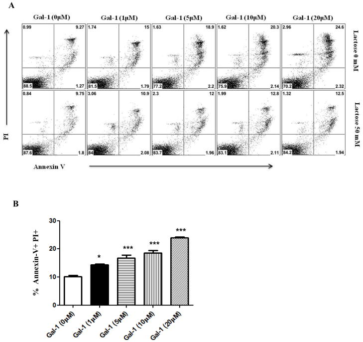
Figure 9. Gal-1 promotes apoptosis of CD4 T cells.
Cells isolated from draining lymph nodes of HSV-1 infected mice (day 15 post infection) were incubated without gal-1 (control) and different concentrations of gal-1, with or without 50mM lactose for a period of 5 hours. After gal-1 exposure, cells were stained with PI, FITC-annexin V and APC- CD4. A–B. Plots shown were gated on CD4+ T cells (A) Representative histograms show Annexin V+ PI+ CD4+ T cells (B) Percentage of Annexin V+ PI+ CD4+ T cells. The experiments were repeated two times. The level of significance was determined using one-way anova using Turkey’s multiple comparison test. Error bars represent mean ± S.E.M.

