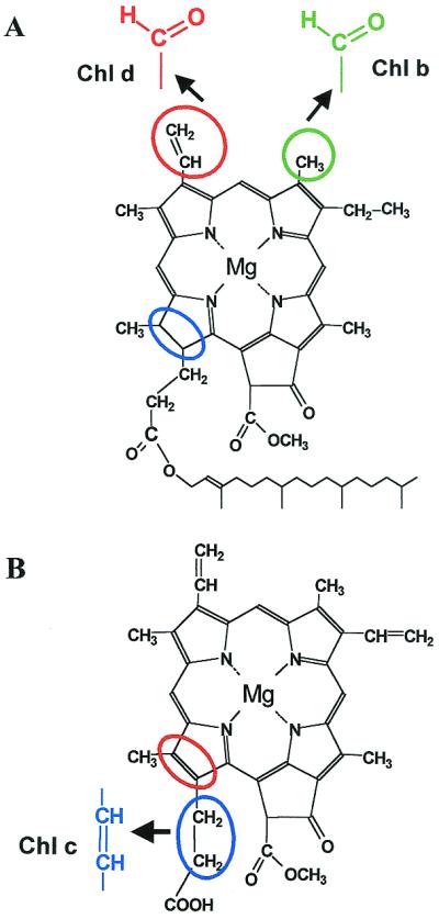
Figure 2.

Structures of eukaryotic Chls. (A) Chlorophyll a. Note the reduced ring D (blue circle) and phytyl tail (not drawn to scale). Side-chain modifications resulting in Chl b (green) and Chl d (red) are shown. (B) Mg-divinyl protochlorophyllide a and Chl c2 (blue). Note the double bond in ring D (red circle).
