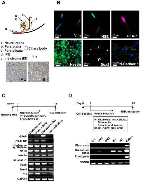
Figure 1. Retinal glia- and retinal progenitor-like phenotypes in iris cells.
(A) Scheme of cell sources in the iris and ciliary body. (B) Immunocytochemical analysis of iris cells. Iris cells are immunocytochemically positive for glial cell marker (GFAP), and neural stem cell markers (Nestin (green), Sox2 (yellow) and N-Cadherin (green)). Nuclei were stained with DAPI (blue) with vimentin, nestin and N-cadherin. (C) Expression of neuron-related genes after neural induction. RT-PCR analysis indicates that iris cells expressed glial cell markers (GFAP, CRALBP and glutamine synthetase), and neural stem cell markers (Nestin, Musashi-1 and Pax6). By the “hanging-drop” method coupled with the B27 medium, rhodopsin was not induced. In this illustration, “Induced” indicates “cells at an induced state by the hanging-drop method coupled with the B27 medium” and “Retina” indicates retina-derived cells at passage 3. (D) Expression of the opsin genes after retinal induction. In this illustration, “w/o” indicates iris-stromal cells without any induction. “IPE” and “IS” indicate “iris pigment epithelial cells” and “iris-stromal cells”, respectively, that were induced by exogeneously added chemicals and growth factors as indicated. By retinal induction with the R1 medium, green/red opsin was up-regulated significantly in iris-stromal cells, but blue opsin or rhodopsin was not up-regulated.

