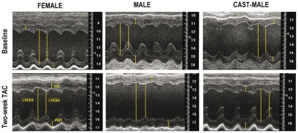
Figure 1. Parasternal short-axis M-mode tracings of the left ventricle obtained from representative mice of each experimental group, before (baseline) and two weeks after aortic constriction (TAC).
LVESd: Left ventricular end-systolic dimension; LVEDd: Left ventricular end-diastolic dimension; PWT: posterior wall thickness; IVST: interventricular septum thickness.

