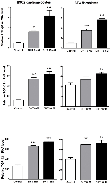
Figure 7. Effects of androgens on TGF-β mRNA expression in cultured H9C2 cardiomyocytes and NIH3T3 fibroblasts.
Cells were exposed to dihydrotestosterone (DHT) for 16 h. Gene expression, determined by real-time PCR, is expressed as the fold induction. Data are the means ± S.E. of three experiments performed in duplicate. *p<0.05, ***p<0.001 vs. control cells (vehicle treated) (ANOVA and Bonferroni post-hoc test).

