FIGURE 4:
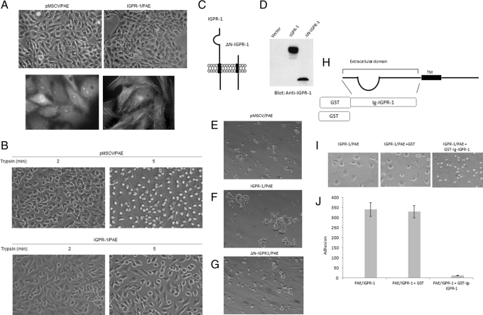
IGPR-1 regulates cellular morphology and adhesion. Morphology of PAE cells expressing IGPR-1 and PAE cells expressing empty vector. Pictures were taken under light microscopy (top). (A) PAE cells expressing empty vector or IGPR-1 were stained with FITC-labeled phalloidin, and pictures were taken under immunofluorescence microscopy. (B) PAE cells expressing either empty vector or IGPR-1 were trypsinized with 0.05% trypsin/EDTA for indicated times, cells were viewed under the microscope, and pictures were taken with a digital camera. (C) Schematic of IGPR-1 and N-terminus-deleted IGPR-1 ((ΔN-IGPR-1). (D) Expression of IGPR-1 and ΔN-IGPR-1 in PAE cells. (E–G) PAE cells expressing empty vector (pMSCV), IGPR-1, or ΔN-IGPR-1 were subjected to aggregation assay as described in Materials and Methods, and pictures were taken under a light microscope attached to a digital camera. (H) Schematic of generation of recombinant GST-IGPR-1. PAE cells expressing IGPR-1 were incubated either with DMEM medium alone, DMEM plus GST, or DMEM plus GST-N terminus/extracellular domain of IGPR-1. After 15 min of incubation, cells were plated in 24-well plates and allowed to adhere. (I) Pictures of cells were taken after 30 min of incubation in 24-well plates. (J) After 30 min, nonadherent cells were removed, and adherent cells were counted a under microscope (three randomly selected fields were counted in each well) and presented.

