FIGURE 4:
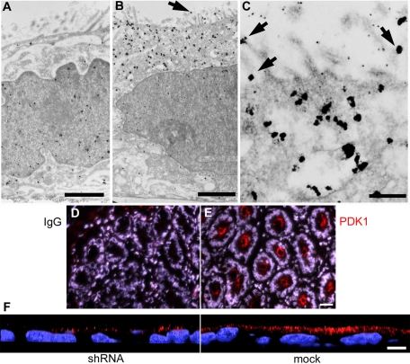
PDK1 distributes to a subapical vesicular compartment and the apical plasma membrane in Caco-2 cells and in intestinal crypts. Cultures were processed for immunogold with (A) nonimmune IgG or (B, C) anti-PDK1 antibody, silver enhanced, and viewed under a TEM. Arrows point at apical membrane-associated gold particles. (D, E) Frozen sections of mouse duodenum were processed with (D) nonimmune IgG, or (E) anti-PDK1 antibody (red channel) for immunofluorescence and counterstained with DAPI (blue channel). (F) Confocal xz-sections of lentiviral PDK1-knockdown Caco-2 cells (shRNA) or mock-transduced cells processed with the same anti-PDK1 antibody (red channel). Bars, A, 1.4 μm; B, 1.9 μm; C, 0.4 μm; D and E, 20 μm; F, 10 μm.

