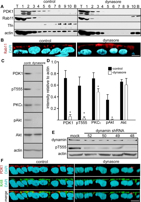
FIGURE 5:
PDK1 comigrates with Rab11 and Tfn in sucrose gradients, and its activity is inhibited by dynasore and dynamin 2 knockdown. (A) The postnuclear supernatants of differentiated Caco-2 cells incubated overnight in Tfn from the apical side and treated with 80 μM dynamin inhibitory peptide dynasore or vehicle only (control) were spun on 10–40% continuous sucrose gradients at 100,000 × g for 20 h. The gradients were fractionated into one sample of the volume seeded on top (T), 10 identical samples of the gradient (1–10), and a wash of the bottom of the tube (pellet, P). The same blots were sequentially reprobed for PDK1, Rab11, Tfn, and actin. (B) The xz reconstructions of confocal stacks of Caco-2 cells grown on filters and treated or not with dynasore were analyzed by immunofluorescence with anti-Rab11 (red channel). (C) Confluent differentiated Caco-2 cells were treated with dynasore or with vehicle DMSO only (contr) in serum-free medium. SDS extracts were analyzed by immunoblot with the antibodies indicated on the left. (D) Quantification of the result shown in C. The bars represent the means ± SD of the ratio of densitometric values of the bands relative to actin bands in the same lane from three independent experiments. For all measurements, nonsaturated images were used (Student's t test significance, *p < 0.005 as compared with the corresponding control). (E) Caco-2 cells were transduced with lentiviral particles with no insert (mock) or four different inserts expressing different shRNAs directed against dynamin 2 (52, 50, 49, 48). SDS extracts were analyzed for immunoblot for dynamin 2, pT555 aPKC, and actin. (F) The xz three-dimensional reconstruction of a confocal stack. Caco-2 cells grown on filters and treated or not with dynasore were analyzed by immunofluorescence with anti-PDK1 (red channel) and anti–keratin 8 antibodies (Krt8, green channel). Bars, 10 μm.

