
Figure 2. Neuronal and glial expression of SFPQ revealed in non-transgenic wild-type (CT) control mouse brain shown for the amygdala (A) and the hippocampus (B,C).
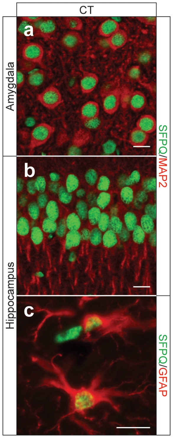
Double immunofluorescence for SFPQ (green)/MAP2 (red) (A,B) and SFPQ (green)/GFAP (red) (C) reveals an exclusively nuclear localization in both neurons and astrocytes of WT mice.