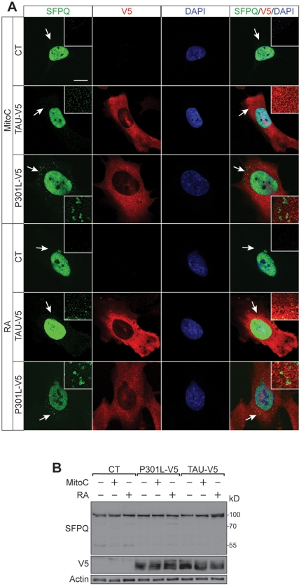
Figure 4. Tau transfection causes SFPQ aggregation in postmitotic cells.
(A) Mitomycin C (Mito C)-mediated cell cycle arrest or neuronal differentiation with retinoic acid (RA) of V5-tagged wild-type or P301L tau-expressing SH-SY5Y compared to untransfected (CT) SH-SY5Y neuroblastoma cells reveals SFPQ aggregates (arrows) in the cytoplasm that are not seen in CT. Insets: detailed view of vesicular SFPQ in the cytoplasm. Nuclear staining: DAPI (blue). (B) Western blotting reveals that total levels of SFPQ are not altered under any of these conditions. Actin has been used for normalisation.

