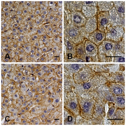
Figure 3. Immunohistochemistry for NMMHC-IIA in the liver biopsy from one patient with MYH9-RD.
Liver biopsy from a 10-years-old patient with MYH9-RD caused by the p.T1155A mutation of MYH9 with persistently elevated AST, ALT and GGT. (A, B): Immunohistochemistry for NMMHC-IIA showed a signal (brown, horseradish peroxidase staining) concentrated close to the hepatocytes' plasma membrane. The distribution of NMMHC-IIA was not significantly different from that from a healthy control (C, D). Specimens were counterstained with Meyer's haematoxylin. Scale bars correspond to 10 µm.

