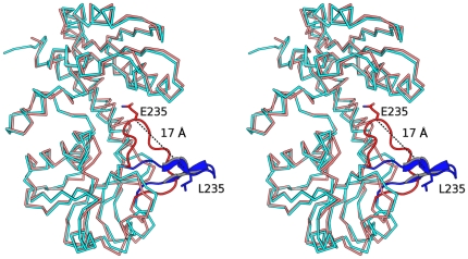
Figure 3. Different loop conformations at the active site.
Superposition of a subunit of E.coli FolD (PDB code: 1B0A black) against PaFolD. A loop in the PaFolD structure (red residues 231–243) adopts a different orientation compared to the EcFolD structure (blue) with equivalent residues (Gln235 Pa and Leu235 Ec) shifting by as much as 16.7 Å and an angle of nearly 60°. In the orientation seen for the PaFolD structure, the loop sits over the active site.

