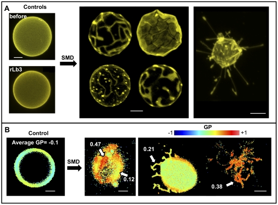
Figure 10. Representative fluorescence microscopy images of the action of SMD on C12SM giant unilamellar vesicles.
A) DiIC18-labeled GUVs. Top left shows a typical vesicle before SMD addition and bottom left a GUV after 17 hours of incubation with inactive Lb3. The center panel shows domain formation in several GUVs 1–3 hours after SMD addition. On the right a collapsed vesicle with extruded tubes. B) Changes in LAURDAN GP induced by SMD. The left panel shows an untreated GUV, homogeneously fluid. The center panel shows domains of different GP value (indicated by arrows). The right panel shows a GUV with tubular extrusions (left) and a collapsed one (right). Bars are 5 µm.

