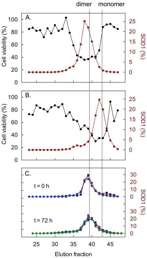
Figure 2. The apoSOD1 molecules do not self-assemble in the cell culture media.
Dimeric and monomeric apoSOD (25 µM) were applied to a Superdex 75 column and the eluted fractions were tested for cytotoxicity in SH-SY5Y cells (black) and analysed for SOD1 by western immunoblotting (red). Cell viability was measured with the resazurin assay and presented as percentage viability of buffer control. The peak areas from the western blot analysis have been normalised to total amount of SOD1 in the chromatography. Cytotoxicity coincides with the dimeric (A) and monomeric (B) peaks of the chromatograms. (C) Analysis of the cell-culture medium directly after protein addition of dimeric apoSOD1 and after 72 h incubation confirms that the apoSOD1 molecules remain dimeric throughout the toxicity assay. Large molecules and aggregates would elute in the void volume of the Superdex-75 column around fraction 26. The concentration of SOD1 in the culture medium was varied between 0.16 µM (black), 0.625 µM (purple), 1.25 µM (red), 2.5 µM (blue) and 12.5 µM (green).

