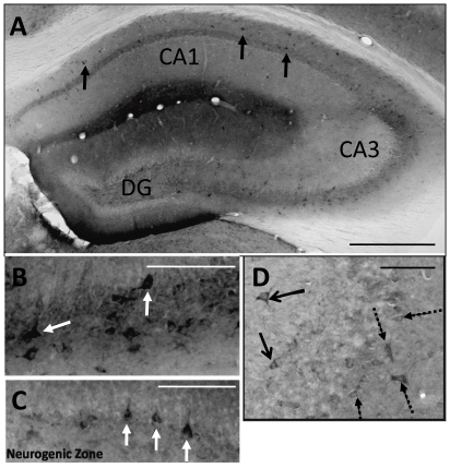
Figure 1. GFP+ cells found in the hippocampus of a 16 week disomic control brain.
A. In this disomic brain, robust mNPC survival was observed with GFP+ cells found in throughout the hippocampus (black cells, arrows). B. mNPC found in the pyramidal layer of CA3 had the typical pyramidal neuronal shape (arrows). C. In the DG, mNPC were found in the subgranule zone, which is the neurogenic layer of the hippocampus (arrows). D. Double labeling for reelin revealed that some GFP+ cells found outside of the pyramidal layer of CA3 were reelin+ interneurons (solid arrows). Dashed arrows indicate host reelin+ interneurons. Scale bars in A = 500 µm and in B–D = 100 µm.

