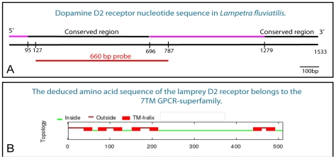
Figure 1. The lamprey dopamine D2 receptor.
A. A schematic drawing of the lamprey dopamine D2 receptor nucleotide sequence showing the conserved regions (black) at the 5′ and 3′ ends, respectively, and the site from which the riboprobe (660 bp, red) was made. B. The seven transmembrane domains (TM-helix) are all located at the conserved parts of the dopamine D2 receptor. The deduced amino acid sequence of the receptor shows that it belongs to the GPCR-superfamily.

