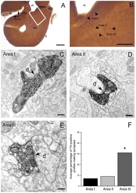
Figure 6. Tyrosine hydroxylase (TH) immunoreactivity in the lamprey striatum.
A and B. Light microscope photomicrographs showing the areas (I, II and III) of the striatum that were included in the electron microscope analysis. Note the slightly higher density of TH-immunoreactive fibers (some indicated by small arrows) in Areas I and III. C–E. Electron micrographs of TH-immunolabeled axonal profiles forming asymmetrical synapses (arrows) with dendritic shafts (d) in Area I (C), II (D) and III (E) of the lamprey striatum. Note the densely packed vesicles and the prominent postsynaptic densities. F. Quantitative analysis of synaptic incidence in the three areas of striatum. The histogram shows the average percentage of TH-immunolabeled profiles which form synaptic junctions (out of a total of 75 profiles per area, n = 3). Synaptic incidence in Area III was significantly higher when compared to Area I (x 2 test, p = 0.0003) and Area II (x 2 test, p = 0.0022). Abbreviations: vt, ventriculus medius telencephali; lpv, ventral part of the lateral pallium. Scale bars: A: 200 µm, B: 100 µm, C–E: 200 nm.

