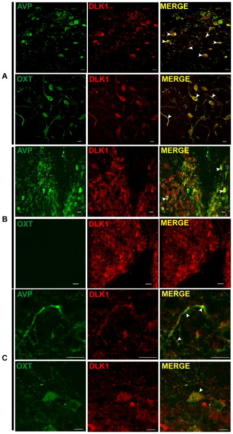
Figure 5. DLK1 expression in AVP and OXT neurons in the PVN, the SCN and the SON.
Dual immunofluorescence staining of DLK1 (antibody C-19) and AVP or OXT on 0.6 µm-thick confocal PVN (A), SCN (B) and SON (C) sections obtained using a 63× objective. Arrowheads indicate double staining. Scale bars: 10 µm.

