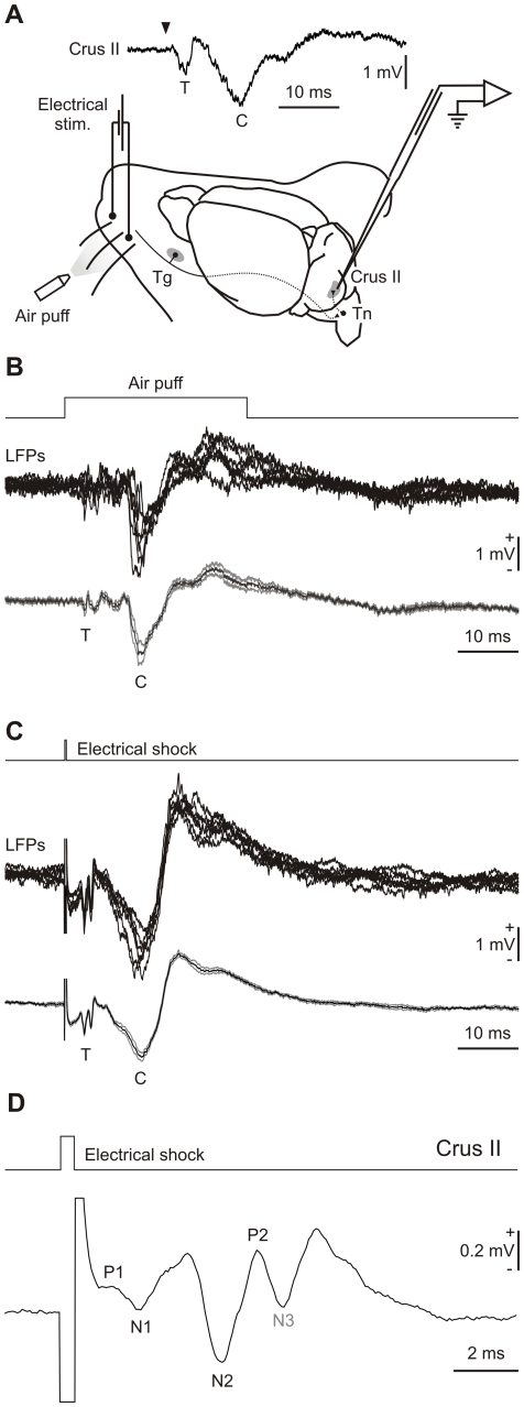
Figure 1. Experimental design and electrophysiological response to electrical stimulation of mouse whiskers.
(A) Animals were prepared for chronic recordings of local field potentials and unitary extracellular activity in the Purkinje cell layer of the Crus I/II area. Facial dermatomes of the whisker region were electrically or tactilely stimulated with a pair of needles under the skin (Stim.) or an air puff pulse, respectively. Sensory information comes into the Crus I/II area from the trigeminal nucleus (Tn) in the brainstem, which receives afferent signals from the trigeminal ganglion (Tg). The LFP recorded in the alert animal induced by tactile stimuli consisted of two major negative waves corresponding to trigeminal (T) and cortical (C) responses (upper trace). (B) In some of the recordings, the T component appeared as two separate components (N2 and N3). The figure shows superimposed LFPs (upper trace, n = 7) and the corresponding mean average (with error bars in gray at bottom) for the LFP acquired after tactile stimulation of the whisker. (C) Superimposed LFPs (upper trace, n = 7) and the corresponding mean average (with error bars in gray at bottom) for the LFP acquired at the same recording place shown in B but after electrical stimulation of the whisker. A major reproducibility of the T-related components in the superimposed traces and a decrement in the error bars in the mean average of LFP were observed after electrical stimulation in comparison to air puff stimulation. (D) The lower trace shows T-related components enlarged from a representative local field potential recording from the Crus II area in the Purkinje cell layer in response to a single-pulse electrical stimulation of the whisker pad (Stim. trace). Early response associated with sensory input in the cerebellum via the trigeminal nucleus is characterized by P1-N1-N2-P2-N3 components.

