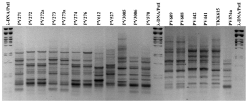
Figure 1. Agarose gel electrophoresis of BOX-PCR of 18 local P. viridiflava isolates.
Agarose gel electrophoresis of BOX-PCR amplification products from genomic DNA of 18 local P. viridiflava isolates. The molecular size marker is λ phage DNA digested with the restriction endonuclease PstI. The negative film filter was applied to the image of an ethidium bromide gel. Isolate codes are given over each lane.

