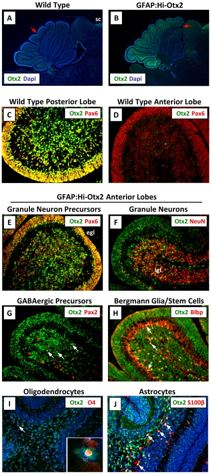
Figure 1. Otx2 expression is induced in various cell types in GFAP:Hi-Otx2 mice.
Sections from P7 wild type (A, C–D) or GFAP:Hi-Otx2 (B, E–J) mice were immunostained with the indicated antibodies. For (A) and (B), anterior expression threshold is designated with a red arrow. (A, B) are shown at 4× magnification (mag), all others 20× mag. sc, superior colliculus; egl, external granule layer; igl, internal granule layer. White arrows indicate overlapping expression of the indicated markers in individual cells.

