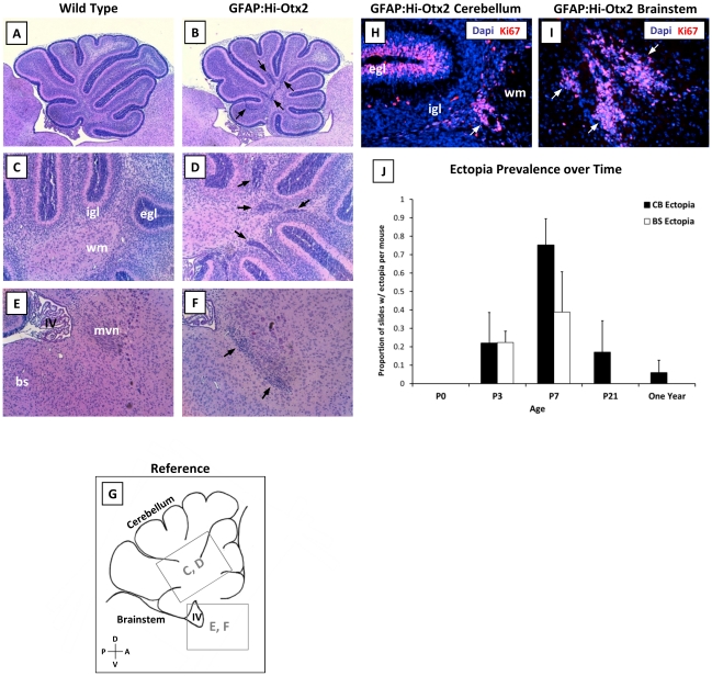
Figure 2. GFAP:Hi-Otx2 mice develop focal hyperplasias in the cerebellar white matter and the brainstem.
(A–F) H & E stained sections from P7 hindbrains of (A, C, E) wild type and (B, D, F) GFAP:Hi-Otx2 mice. Fields shown are as follows: (A, B) whole cerebella at 4× magnification (mag), (C, D) cerebellar white matter at 10× mag, (E, F) dorsal brainstem at 10× mag. (G) reference illustration of fields shown in C–F indicated by grey boxes. (H, I) Immunofluorescent staining for Ki67 in (H) cerebellar ectopia and (I) brainstem ectopia in GFAP:Hi-Otx2 mice (20× mag). (J) Prevalence of ectopia in GFAP:Hi-Otx2 mice over time. CB, cerebellum; BS/bs, brainstem; egl, external granule layer; igl, internal granule layer; wm, white matter; IV, fourth ventricle; mvn, medial vestibular nuclei; D, dorsal; V, ventral; A, anterior; P, posterior. Black or white arrows indicate ectopia.

