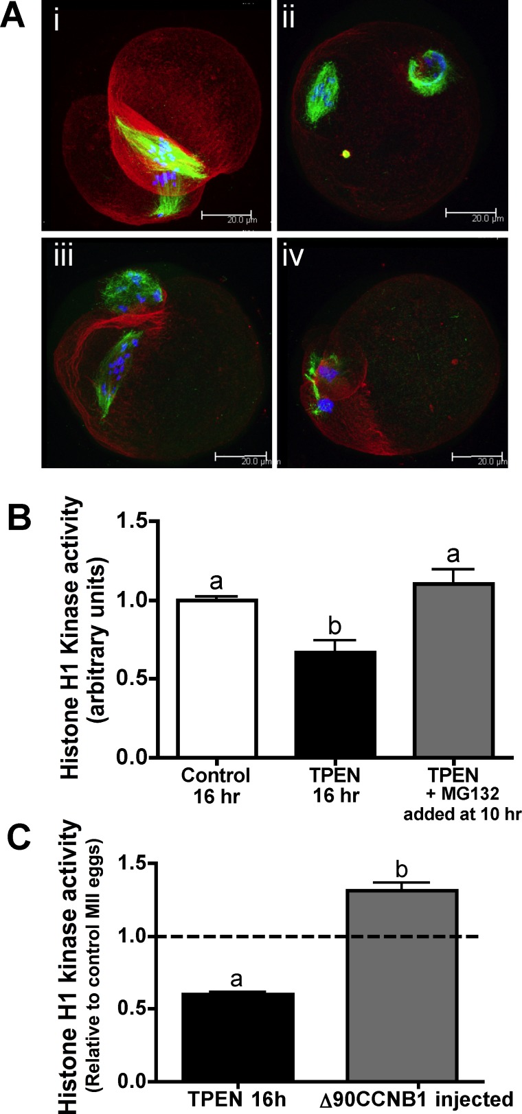
FIG. 2. .
Restoration of MPF activity following the MI-MII transition partially rescues zinc-insufficient oocytes. A) COCs were cultured in TPEN-containing medium for 10 h and transferred to medium containing both TPEN and MG132 for an additional 6 h followed by removal of cumulus cells. Spindle stains show that 69% of oocytes had some degree of MII spindle formation, with 19% showing aligned metaphase plates (i), 31% showing three or fewer misaligned chromosomes (ii), and 19% showing more than three misaligned chromosomes (iii). A majority of oocytes cultured in TPEN without MG132 for the entire culture period displayed the TI-arrested spindles associated with zinc insufficiency during IVM (iv). (Refer also to Supplemental Table S2.) Three-dimensional projections of confocal Z stacks with actin (red), tubulin (green), and DAPI (blue) are shown. Bar = 20 μm. B) Graph of histone H1 kinase activity showing densitometric analysis for at least six individual oocytes per group. Error bars represent the SEM, and different letters indicate significant differences according to ANOVA with Bonferroni post-hoc test (P < 0.01). C) Histone H1 kinase activity for oocytes cultured in TPEN containing medium for 10 to 12.5 h followed by injection with CCNB1(Δ90)-EGFP cRNA and transfer back to TPEN containing medium for 3.5 to 6 h is presented as in B.

