FIG. 4. .
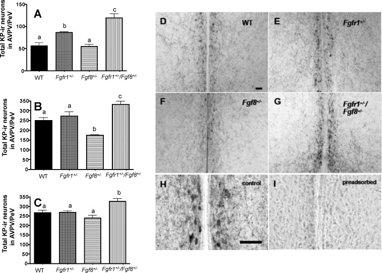
Total AVPV/PeV KP-ir neurons in WT, Fgfr1+/−, Fgf8+/−, and Fgfr1+/−/Fgf8+/− mice on PND 15 (A), PND 30 (B), and PND 60 (C). Different letters represent significant differences (P < 0.05). n = 4–6 for all age groups. D–G) Representative photomicrographs of KP-ir neurons in PND 30 WT (D), Fgfr1+/;− (E), Fgf8+/− (F), and Fgfr1+/−/Fgf8+/− (G) female mice. H, I) Representative images of adjacent sections incubated with KP antibody that has not (H) or has (I) been preadsorbed with 200 μg/ml KP10. Bar in D applies to D–G; bar in H applies to H and I. Bars = 50 μm.

