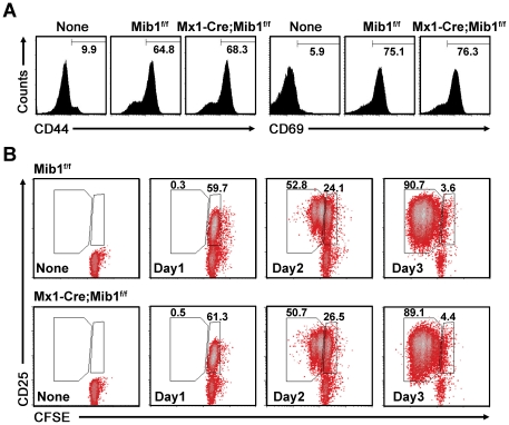
Figure 4. Preserved activation and proliferation of Notch-inactivated CD4+ T cells.
A, Flow cytometry analysis for T cell activation markers, CD44 and CD69, in purified CD4+ T cells before (None) or 24 h after coculture with Mib1f/f or Mx1-Cre;Mib1f/f DCs. B, CFSE-labeled naive OT-II CD4+ T cells were cultured without (None) or with peptide-pretreated Mib1f/f or Mx1-Cre;Mib1f/f DCs, and the activated cells collected on days 1, 2, and 3, respectively, were stained for CD25 and CD4 and analyzed by flow cytometry. The numbers indicate the percentage of cells within the gates. A representative of three independent experiments is shown.

