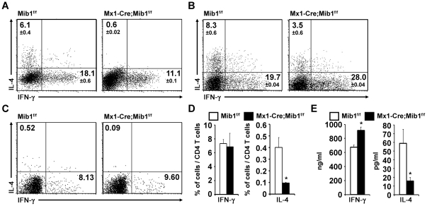
Figure 6. Mib1 in DCs is critical for Th2 induction.
A, Neutralizing anti-IFN-γ Abs (10 µg/ml) were treated to CD4+ T cells during the coculture with peptide pretreated Mib1f/f or Mx1-Cre;Mib1f/f DCs. Intracellular cytokine expression was measured by flow cytometry, as shown in Figure 5A . The numbers indicate the mean ± SD cell percentages from two independent experiments. B, Purified naïve SMARTA CD4+ T cells were stimulated with peptide-pretreated Mib1f/f or Mx1-Cre;Mib1f/f DCs. Intracellular cytokine expression was measured by flow cytometry, as shown in Figure 5A . The numbers indicate the mean ± SD cell percentages from two independent experiments. C, Purified naive OT-II CD4+ T cells were transferred intravenously into CD45.1 recipient mice. Peptide-pretreated Mib1f/f or Mx1-Cre;Mib1f/f DCs were intraperitoneally injected on the subsequent day. Seven days later, splenocytes were re-stimulated for 6 h with OVA323–339 peptides and human rIL-2, and intracellular IFN-γ and IL-4 were analyzed from CD45.2+ CD4+ cells by flow cytometry. The numbers indicate the percentage of cells within the gates. A representative of three independent experiments is shown. D, The average percentage of activated IFN-γ and IL-4-producing CD45.2+ CD4+ T cells (as shown in [C]) from three independent experiments. E, IFN-γ and IL-4 productions were detected by ELISA 72 h after re-stimulation. *, P<0.05.

