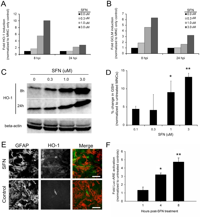
Figure 5. Sulforaphane elevates HO-1 expression via Nrf2/ARE activation in astrocytes.
SFN (0, 0.3, 1.0 or 3.0 µM) was added onto MNC for 8 & 24 h prior to collection for semi-quantitative RT-PCR (A & B) and Western blot (C) analysis. RT-PCR of HO-1 (A) and GCLM (B) mRNA expression in MNCs following 8 & 24 h exposure to SFN. Data are presented as fold induction over saline controls and are representative of 3 separate experiments. (C) Western blot analysis of HO-1 protein expression in MNCs following 8 & 24 h exposure to SFN. (D) GSH concentration in MNCs following 48 h exposure to SFN (p≤0.0001). (E) SFN treated MNC (24 h) stained for HO-1 (green) and GFAP (red). Scale = 50 µm. (F) ARE-Luciferase reporter assay in purified astrocyte cultures following 4 and 8 h SFN treatment. *p = 0.007; **p = 0.006. Statistical significance was tested using an ANOVA single factor with PLSD Post-hoc analysis (StatView 5).

