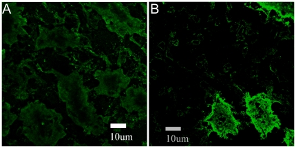
Figure 7. Sample CLSM images of native NBCe1-A on basolateral cell membranes of rat kidney.
Post fixation, tissues were immunolabeled with the primary anti-wt-NBCe1-A antibody (I) and the secondary (II) Alexa Fluor 488 conjugated antibody. Panel A shows non-specific secondary antibody staining in the absence of the primary. Panel B shows kidney cells immunostained with both antibodies. The pixel size is 0.0921 µm. The images were taken at identical acquisition conditions so valid comparisons between different images could be made. The image contrast was enhanced for visualization and comparison purposes.

