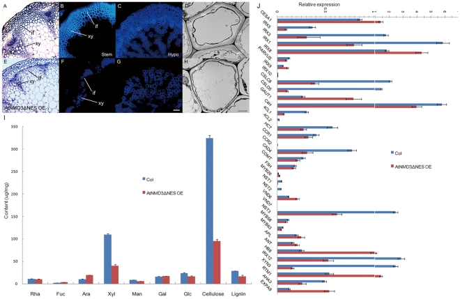
Figure 5. Secondary cell wall thickening is severely defective in the AtNMD3-ΔNES OE line.
A to H show the defect of secondary cell wall thickening in the AtNMD3ΔNES OE line. A to D show normal morphology of the Col-0 stem after semi-thin plastic sectioning (A) and free-hand sectioning for UV observation of the secondary cell walls (B), of Col-0 hypocotyl after free-hand sectioning (C), and of Col-0 stem after ultra-thin sectioning for TEM observation (D). E to H show abnormal morphology of counterpart samples collected from the AtNMD3ΔNES OE lines, indicating the defect of the secondary cell wall thickening. I shows the quantification of the cell wall components, indicating dramatic reductions of xylan (Xyl), cellulose and lignin. J shows the expression levels of cell wall related genes. (if: interfascicular fiber; xy: xylem; SW: secondary cell wall. Bar for A, B, C, E, F, G = 50 µm; D, H = 2 µm).

