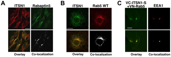
Figure 3. ITSN1 interacts with components of the Rab5 GTPase pathway.
A. ITSN1 interacts with Rabaptin5. Endogenous ITSN1 (red) in HeLa cells co-localizes with myc-tagged Rabaptin5 (green). Antibodies to Rabaptin5 did not detect the endogenous protein. Overlap of proteins is shown in yellow. Regions of overlap were extracted from the image using ImageJ. B. Endogenous ITSN1 (green) co-localizes with endogenous Rab5 (red) in COS cells. Overlap of proteins is shown in yellow. Regions of overlap were extracted from the image using ImageJ. C. ITSN1-S forms a complex with Rab5 on early endosomes. HeLa cells were transfected with VN-Rab5 and VC-ITSN1-S and then stained with antibody to EEA-1 (red). The BiFC complex of Rab5 and ITSN1-S is pseudocolored green. Overlap of the BiFC complex with EEA1 is shown in yellow. Regions of overlap were extracted from the image using ImageJ. Note: white scale bars represent 20? m. The fluorescence patterns shown in all panels are representative of localization observed throughout the plate.

