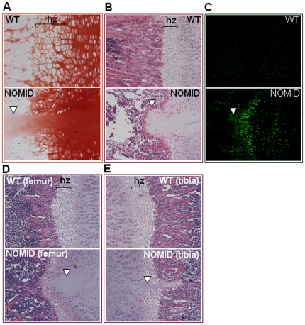
Figure 4. NOMID mice exhibit disorganized growth plates.
Femoral sections from P13 (A–C) or P8 (D and E) mice were used for safranin O (A) and H&E (B, D and E) staining or for TUNEL (C). Original magnification: ×20 (A and C), ×10 (B, D and E). The spike (arrowhead) and early morphological changes (D, arrowhead) were observed only in NOMID mice. NOMID cells showed a high degree of apoptosis. hz, hypertrophic zone.

