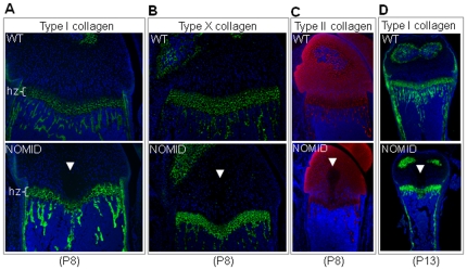
Figure 5. Type II collagen staining is reduced in the acellular structure in NOMID mice.
Femoral sections from P8 (A, B and C) or P13 mice (D) were stained for types I, X (green), II (red) collagen, and counterstained with DAPI (blue). Type II collagen staining was not observed in the acellular structure within the cartilage zone above the hypertrophic chondrocyte zone (hz). Original magnification: ×20 (A and B), ×10 (C and D).

