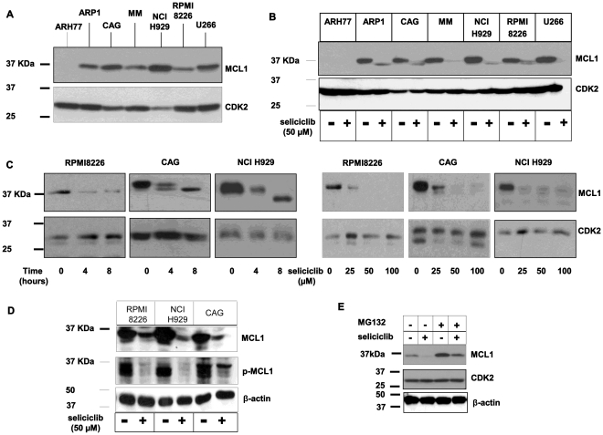
Figure 4. Seliciclib downregulates MCL1 expression in hMMCLs.
(A) The indicated multiple myeloma cell lines in logarithmic growth phase were extracted and subjected to immunoblotting, utilizing MCL1 antibodies. In all experiments CDK2 expression serves as an internal loading control. Experiments were performed at least 3 times and one representative result is presented. (B–C) MCL1 downregulation by seliciclib. Cells were extracted and subjected to immunoblotting. (B) Various hMMCLs were incubated in the presence of seliciclib 50 µM or DMSO for 6 hours and the level of MCL1 expression was analyzed. (C) RPMI8226, CAG and NCI H929 cells were incubated in the absence or presence of seliciclib 50 µM for the indicated time points, or in the presence of increasing concentrations of seliciclib for 8 hours. (D) Reduction in MCL1 phosphorylation by seliciclib. Cells were treated with 50 µM or DMSO for 8 hours and the levels of total and phosphorylated MCL1 were analyzed by immunoblotting using specific antibodies. β-actin was used to confirm equal protein loading. (E) CAG cells were incubated in the absence or presence of seliciclib or MG132 (10 µM) exclusively or combined. MCL1 level of expression was verified by immunoblotting. β-actin was used to confirm equal protein loading.

