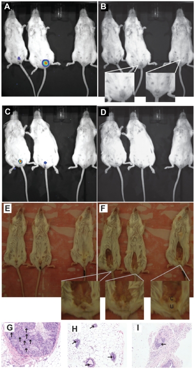
Figure 3. Infection localization.
A) Mice infected with bioluminescent B. melitensis GR023 shown after luminescence imaging. B) The same image as (A) with the bioluminescence removed to show edema in the urogenital area. The uninfected mouse is on the right side of the frame. A close up of the infected mouse with edema and the control mouse are shown. Mice infected for 450 days are shown with C) bioluminescence, D) with bioluminescence removed, E) just prior to dissection of urogenital area, and F) after dissection showing the clitoral gland (c) just subcutaneous of the ureter (u). Female BALB/c mice infected for 240 days at 20× magnification with infiltration of neutrophils, lymphocytes and macrophages shown by arrows in the G) interstitial region of the clitoral gland, H) lumen of the mammary glands, and I) perivascular cuffing in the subcutaneous tissue of the perianal area.

