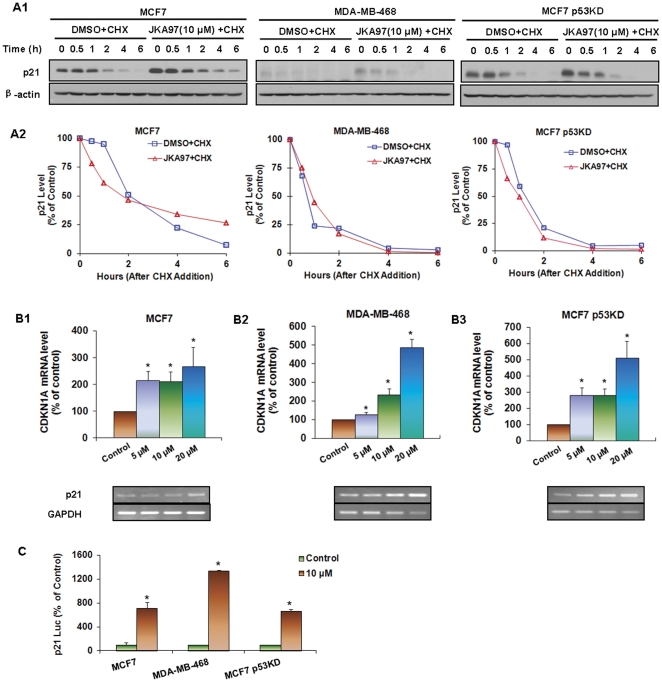
Figure 4. Effects of JKA97 on p21 expression.
(A1) MCF7, MDA-MB-468, and MCF7 p53KD cells were exposed to various concentrations of JKA97 or vehicle for 24 hrs, followed by exposure to protein synthesis inhibitor cycloheximide (CHX, 10 µg/mL). p21 protein expression was detected by Western blotting at different times after exposure of CHX. (A2) The graph shows the quantification of the Western blotting data. MCF7 (B1), MDA-MB-468 (B2) and MCF7 p53KD (B3) Cells were exposed to various concentrations of JKA97 or vehicle for 24 hrs, and total RNA were extracted followed by reverse transcription, and detection mRNA level of p21 by Real-time quantification PCR and Quantification RT-PCR, normalized by mRNA level of GAPDH. (C) Cells were transfected with p21 promoter luciferase reporter plasmid and a Renilla luciferase reporter together for 12 hrs, followed by treatment of 10 µM JKA97 or vehicle for an additional 24 hrs. The reporter activity was normalized to the corresponding Renilla luciferase reporter. The luciferase assay was performed in triplicate. Statistical significance was determined compared with control (*P<0.05).

